Ultrasound
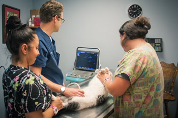
Although humans and animals are different in many ways, some advances in human medicine are also very useful for veterinary patients. One of these advances, diagnostic ultrasound, has proven to be a powerful tool in veterinary medicine. At Shelby Center Hospital for Animals, one of our goals is to offer state-of-the-art medicine and diagnostic testing. SCHA is pleased to offer ultrasound services as a means of providing a higher level of quality care to our patients.
Ultrasonography is a type of diagnostic technique that uses ultrasound waves to produce an imaging study. This means that when we perform ultrasonography, we can see internal images of the patient’s body. Unlike some other imaging studies, like x-rays, ultrasonography uses a minute dose of radiation that is nearly undetectable. Ultrasonography uses high-frequency sound (ultrasound) waves to create a picture of what is inside your pet’s body. Ultrasonography is a completely non-invasive, painless way to diagnose and evaluate many common diseases.
The ultrasound procedure requires the use of a special machine that emits pulses of ultrasonic waves into soft tissue. These ultrasonic waves bounce off of organs, soft tissue and masses in your pet’s body and return to a sensor inside the ultrasound machine. The ultrasound equipment collects these reflected “echoes” and uses them to generate images that are viewable on a screen.
Ultrasound waves can generate excellent images of abdominal organs, including the liver, spleen, gallbladder, and kidneys. It is also useful for assessing fetal health and monitoring pregnancy in breeding animals, and it can help us diagnose and stage (determine the severity of) some forms of cancer. The heart can also be evaluated using ultrasound. Heart function, valve defects, clot identification and heart wall damage can all be found using ultrasound. Shelby Center Hospital for Animals is pleased to offer this state of the art service for its clients.